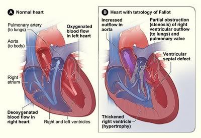
What Is It?Tetralogy of Fallot occurs in about 5 out of every 10,000 babies. There is no known reason for this. It just happens. Tetralogy of Fallot has four key features.
Large Ventricular Septal Defect (VSD) - A VSD is a hole in the part of the septum that separates the ventricles—the lower chambers of the heart. The hole allows oxygen‑rich blood to flow from the left ventricle into the right ventricle instead of flowing into the aorta, the main artery leading out to the body.
Pulmonary Stenosis - This is a narrowing of the pulmonary valve and the passageway through which blood flows from the right ventricle to the pulmonary arteries. Normally, oxygen-poor blood from the right ventricle flows through the pulmonary valve into the pulmonary arteries and out to the lungs to pick up oxygen. In pulmonary stenosis, the heart has to work harder than normal to pump blood, and not enough blood can get to the lungs.
Right Ventricular Hypertrophy - This is when the right ventricle thickens because the heart has to pump harder than it should to move blood through the narrowed pulmonary valve.
Overriding Aorta - This is a defect in the location of the aorta. In a healthy heart, the aorta is attached to the left ventricle, allowing only oxygen-rich blood to go to the body. In tetralogy of Fallot, the aorta is between the left and right ventricles, directly over the VSD. As a result, oxygen‑poor blood from the right ventricle can flow directly into the aorta instead of into the pulmonary artery to the lungs.Together, these four defects mean that not enough blood is able to reach the lungs to get oxygen, and oxygen-poor blood flows out to the body. So TOF does not mean the heart is failing, it really means the child is suffocating. Babies and children with Tetralogy of Fallot have episodes of cyanosis (si-a-NO-sis), which is a bluish tint to the skin, lips, and fingernails. Cyanosis occurs because the oxygen level in the blood is below normal. Babies and children can also have what are called TET spells or blue spells when their oxygen levels get too low and the child may quit breathing. Ever heard of a "blue baby" if so, this is what they were referring to. For this reason, the child must not get too upset, crying can easily bring on a tet spell. Tetralogy of Fallot must be repaired with open-heart surgery, either soon after birth or later in infancy.
Complete Repair -To do a complete repair, the surgeon closes the ventricular septal defect with a patch and opens the right ventricular outflow tract by removing some thickened muscle below the pulmonary valve, repairing or removing the pulmonary valve and enlarging the peripheral pulmonary arteries that go to both lungs. Sometimes a tube is placed between the right ventricle and the pulmonary artery.
